Post-op treatment of anterior cruciate ligament tears
The clinic receives a visit from a 9-year-old spayed female working line German Shepherd with a 7/9 body condition following anterior cruciate ligament tear surgery and subsequent removal of one of the pins that could be uncomfortable for the patient due to an intra-articular position.
The medical history shows a mixed diet of kibble and BARF. Two months since the surgery, the patient continues to present 5/5 limping of the rear right leg with no improvement. A bone biopsy is performed with no significant findings and anti-inflammatory drugs are taken for a long period of time.
Clinical situation of the patient
A left-shifted centre of gravity is observed while at rest during the functional assessment and the patient presents limited capacity to flex the right knee in the seated position. When walking, the patient presents 5/5 limping of the rear right leg (Video: Lola first session), as well as increased lumbosacral movement on the vertical plane.
An increased surface temperature can be felt when touching the right knee, with noticeable atrophy of the leg and hypertrophy of the contralateral limb. Upon specific muscular palpation, significant atrophy of the affected limb is observed, hypertrophy of the contralateral extremity, paravertebral muscle pain at the lumbar level, pain in the left iliopsoas, an overloaded thoracic trapezius and both triceps.
Upon specific articular palpation, the patient presents limited flex in the right knee with pain to the touch in the patellar ligament, as well as crepitation.
Crepitation in both carpi, with thickening in the phalanges and crepitation. All other joints feel normal to the touch.
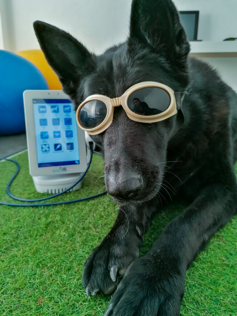
A decision is taken to treat the inflammation with laser, initially the affected knee with the inflammation protocol adapted to suit the area of this patient. Given the handling difficulty, the scanner hand tool is used in non-contact mode and the scanning technique. Treatment is applied twice per week. Controlled activity and diet is also recommended during this first session.
Improvements during laser therapy sessions
At the start of the second session, the patient presents less discomfort upon manipulation so the scanner hand tool is replaced with the massage hand tool in contact mode, maintaining the scanning technique.
At the start of the third session, the patient presents with 3/5 limping – a significant improvement. The same application mode and technique are maintained but the knee protocol is added in order to also treat the contralateral knee and prevent possible injury.
At the fourth session, a decision is made to combine laser therapy with hydrotherapy and massages for the overloads presented by the patient. The same treatment mode and technique continue to be used. The hips are also treated using the massage hand tool. The inflammation protocol is removed from the right knee, maintaining the knee protocol.
The dose is increased to +25% over the course of sessions on the right knee.
At the eleventh session, the patient presents with 1/5 limping. Upon palpation, the patient presents a clear improvement in knee flex, the centre of gravity has improved (albeit not perfect) and general wellbeing has improved.
Hence, laser therapy was useful in this case for treating a two-month limp stemming from anterior cruciate ligament tear surgery.
Do you want to know more about DoctorVet applications?