Moist dermatitis or hot spots is characterised by the presence of erythematous focal injuries anywhere on the body with a moist appearance and partial alopecia. It is important to know the cause of such dermatitis, which might be hypersensitivity to a flea bite, trauma abrasions, behavioural problems or foreign bodies. Excessive licking worsens these lesions1. One of the most common micro-organisms that appears in this kind of dermatitis is Staphylococcusintermedius2.
A superficial and ulcerative inflammatory pattern has been observed at a histological level. Another pattern has been one of suppurative folliculitis; i.e. localised pyoderma.
The diagnosis should include any factors possibly triggering the dermatitis.
Drug treatment and laser therapy for dermatitis in dogs
The treatments include shaving the wounded area and surrounding healthy skin, specific shampoos, topical treatments with both corticosteroids and antibiotics, as well as systemic treatments that include corticosteroids and antibiotics depending on the seriousness of the lesions. Furthermore, other therapeutic options will be included based on the underlying cause3.
The various general effects of laser therapy should be considered in terms of inflammation control4, infection5 and the general effects on dermatology6. Therapeutic laser can be useful as an adjuvant therapy for dermatitis, helping to reduce erythema and re-establishing cutaneous integrity. An antimicrobial effect has been seen against other Staphylococcus7, meaning it can be useful in reducing the amount of antibiotics administered. This can reduce the side effects of systemic treatment.
Photodynamic therapy should be based on combining a certain wavelength with a photosensitising drug. Although more studies in veterinary medicine on the use of photodynamic therapy are needed, it has proved useful in human dermatology8.
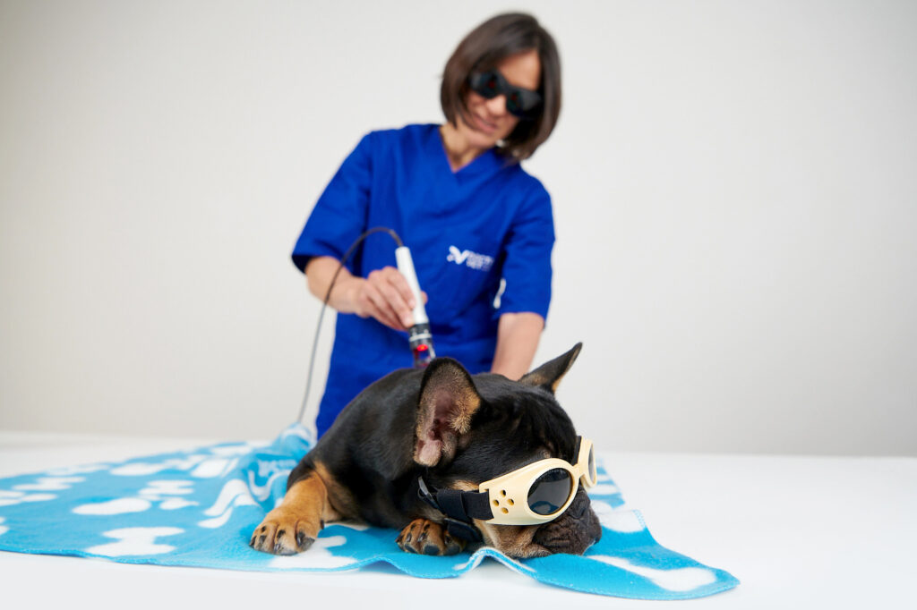
DoctorVet has protocols that can be combined to help the treatment of dermatitis in dogs. Based on the characteristics of the pathology and the histology, the recommended protocols would be those of inflammation, superficial and/or open flesh infection based on the appearance of the lesion, with an assessment by the veterinarian being fundamental.
Furthermore, when dealing with an acute process, we recommend reducing the dose (by 25-50%) according to the combination of various protocols. On the other hand, when dealing with a chronic process and according to the combination of various protocols, those doses can be maintained or increased by 25-50%.
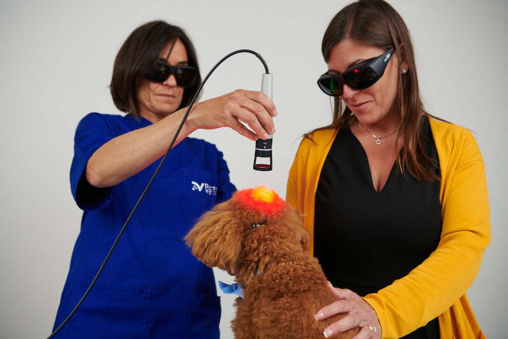
The sweeper treatment head should be used with the scanner application technique in non-contact mode. The treatment schedule may vary between 2-5 sessions/week during the induction stage depending on whether you are dealing with an acute or chronic process. These sessions can be spread further apart once an improvement is observed.
BIBLIOGRAPHY
Palmer LE. Picture This: management of canine pyotraumatic dermatitis (a.k.a, hot spot). J Spec Oper Med, 18(2): 105-109, 2018.
Holm BR et al. A prospective study of the clinical findings, treatment and histopathology of 44 cases of pyotraumatic dermatitis. Vet Dermatolg, 25(3): 369-376, 2004.
Kersey KM. et al. Dermatologic emergencies: identification and treatment. Compend Contin Educ Vet, 35(1), 2013.
Kuffler et al. Photobiomodulation in promoting wound healing: a review. Regen.med. 2015.
Kaya GS. et al. The use of 808-nm light therapy to treat experimental chronic osteomyelitis induced in rats by methicillin-resistetant Staphylococcus aureus. Photomed Laser Surg, 29(6): 405-412, 2011.
Avci P. et al. Low-level laser (light)therapy (LLLT) in skin: stimulating, healing, restoring. Semin Cutan Med Surg, 32(1): 41-52, 2013.
Krespi YP. Et al. Laser-assited nasal decolonization of Syaphylococcus aureus, including methicillin-resistant Staphylococcus aureus. Am J Otolaryngol, 33(5): 572-575, 2012.
Rodenbeck DL. et al. Phototherpay for atopic dermatitis. Clin Dermatol, 34(5), 607-613, 2016.
Do you want to know more about DoctorVet applications?