Pododermatitis, also known as avascular necrosis, is a frequent pathology in the exotic field of speciality. To understand how laser therapy can be useful, it is important to know the anatomy of rabbits and the factors that trigger this pathology.
Rabbits are characterised by a lack of paw pads. The skin is thin and attached to the underlying tissues. Fur helps protect the underside of the metatarsal, rabbits being digitigrade animals. The digital flexor is in constant tension to enable jumping.
Causes and characteristics of pododermatitis in rabbits
Some of the causes of pododermatitis are excess weight or a poor diet, a lack of activity, a cage with inadequate flooring, moisture and a lack of hygiene.
Pododermatitis leads to ligament erosion, leading to poor weight distribution and causing further injury. In turn, this leads to less mobility and even further injury1.
Due to the redistribution of weight owing to the factors that trigger pododermatitis, pressure is applied to the metatarsal area that leads to ischemic inflammation and necrosis of the soft tissues. This can cause thrombosis in small blood vessels. The visible signs include inflammation, erythema and callosity.
Chronic inflammation leads to hyperkeratosis of the epithelium, as well as superficial ulceration. If not treated immediately, the wound can affect subcutaneous tissues and erode the underlying vasculature, leading to haemorrhaging. Furthermore, this wound can be affected by a bacterial infection with the most common microorganisms in rabbits, being S aureus and Pasteurella multocida, as well as fungi1.
Pododermatitis may progress to affect the ligaments, tendons and bone tissue, this stage being the most conservative prognosis. Other complications include septicaemia as a consequence of the infection.
Classification and diagnosis of pododermatitis
Various classifications exist based on the seriousness. The first is Jenkins with a scale of 1-32, while Mancinelli classifies the disease on a scale of 0-63, with 3 and 6, respectively, showing the highest level of wounding with effects on the deepest tissue layers.
A medical history is fundamental for diagnosis, as well as the clinical signs from the patient, a correct examination and possibly the completion of cultures, cytology, x-rays and blood analysis.
Treatment is based on the degree of seriousness and seeks to correct the underlying factors (weight control, diet correction, improved hygiene and mobilising the patient), to treat the wounds themselves and to administer antibiotics and analgesics.
Use of laser therapy to support the treatment of pododermatitis
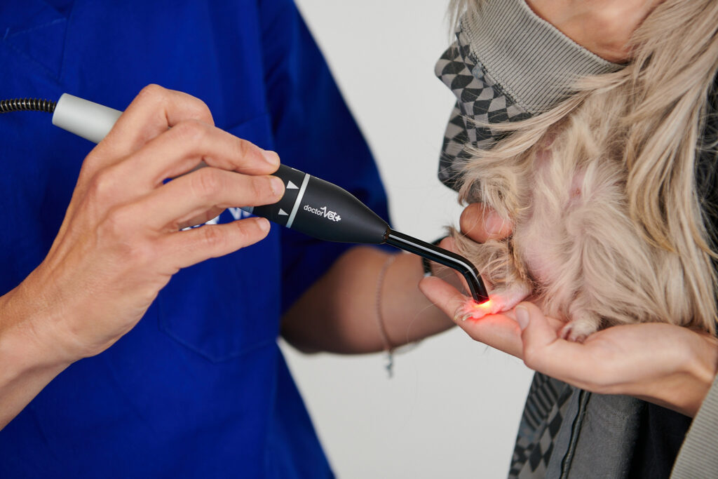
Furthermore, Blair et al. recommend laser therapy to help the healing process, with treatments twice a week for three weeks, as well as multimodal therapy that includes the afore-mentioned treatments2.
Hence, although further study is required, the effects of laser therapy make it an effective tool in the multimodal treatment of pododermatitis in rabbits. It can heal soft tissues, offer benefits in tendinopathies and bone problems and provide an antimicrobial role against S. Aureus4.
Based on the article by Blair, DoctorVet can help to maintain the recommended treatment schedule. In cases where only the soft tissue is affected, the protocols of superficial infection, inflammation and healing can be combined. If deeper tissues are affected, a combination of protocols for deep infection and a choice of articular problems is recommended. The treatment is applied using the scanner hand tool in non-contact mode.
BIBLIOGRAPHY
Varga. Textbook of rabbit medicine. 2nd edition. Butterworth-Heinemann. 2013
Blair et al. Bumblefoot: a comparison of clinical presentation and treatment of pododermatitis in rabbits, rodents, and birds. Vet Clin North Am exot Anim Pract. (3): 715-735. 2013
Mancinelli et al. Husbandry risk factors associated with hock pododermatitis in UK pet rabbits (Oryctolagus cuniculus). Vet. Record 26. 2014
Borstein et al. Near- infrared photoinactivation of bacteria and fungi at physiologic temperatures. Photochem-photobio. (6): 1364-1374. 2009
Do you want to know more about DoctorVet applications?