Otohaematoma is an accumulation of blood between the cartilage of the ear and the skin. This accumulation of blood can affect the entire ear surface or just a part of it. Its precise aetiology is unknown but it seems to be related to head shaking and excessive ear scratching caused by otitis externa1. The haematoma stems from the branch of the main auricular artery within the broken cartilage1.
Diagnosis and treatments
Diagnosis is made by physically examining the patient. A soft fluctuating thickening can be felt initially. With time, this hardens into the classic cauliflower appearance2.
A number of different therapy options exist, including surgery1,2, of which some have already been described with CO2 laser3. Other known options include making an incision and installing drains, stitching along the length of the ear, piercing the ear or the application of compressive bandages1,2.
Laser treatments for otohaematoma
Due to its ability to stimulate micro-circulation4, laser therapy improves the re-absorption of haematomas while also leading to faster fibrin degradation5,6. Laser therapy can be used in combination with the aforementioned therapeutic techniques. Furthermore, it has been shown that laser has a biostimulant effect on cartilage, potentially helping it to heal and shortening recovery times.
DoctorVethas specific protocols that help resolve otohaematoma. A combination of the inflammation, infection and post-surgery protocols is recommended immediately after performing one of the surgical techniques mentioned above.
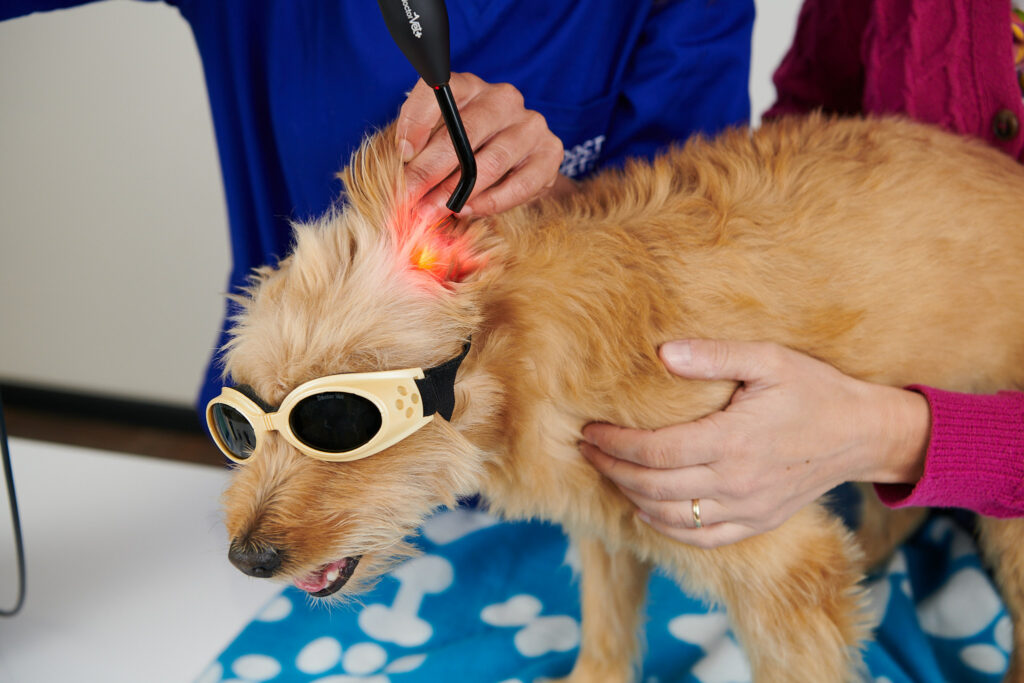
Following this acute post-surgery stage, the inflammation and subcutaneous protocols can be combined. In these cases, we recommend using the sweeper treatment head in non-contact mode and the sweep technique, applying treatment from the base of the ear to the tip.
For cases involving the drainage of that accumulation of blood7, and if the patient can tolerate the procedure, we recommend using the massage treatment head in contact mode and the sweep technique from the base to the tip of the ear. The inflammation control and pressure ulcer protocols are used in this situation. On the other hand, if the patient cannot tolerate contact mode, this can be switched for the non-contact mode and the sweeper hand tool.
The treatment for otitis should be applied when such cases arise as the effects of laser can have a positive impact.
The treatment schedule in these cases is 2-3 times/week. Once an improvement is observed, these sessions can be spread further apart to 1-2 times/week until the problem has been resolved.
BIBLIOGRAPHY
Fossum TW. et al. Cirugía en pequeños animales. Ed. Elseviar, 3ª Edición. Cap. 17: 307-312, 2009.
MacPhail, C. Current treatment options for auricular hematomas. Vet Clin North Am Small Anim Pract, 46(4): 635-641, 2016.
Dye TL et al. Evalutaiton of a technique using the carbon dioxide laser for the treatment of aural hematomas. J Am Anim Hosp Assoc, 38(4): 385-390, 2002.
Karu et al. Cellular effects of low power laser therapy can be mediated by nitric oxide. Lasers in surg. Medicine 36: 307-314 (2005).
Burgudzhieva T. The analgesic and resorptive laser therapy of wound infiltrates, seromas and hematomas in episiorrhaphy and perineorrhaphy. Akush Ginekol, 29(1): 36-39, 1990.
Medrado AP et al. Influence of laser phtobiomodualtion upon connective tissue remodeling during wound healing. J Photochem Photobiol B, 92(3): 144-152, 2008.
Lahiani J. et al. On the nature of canine aural haematoma and its treatment with continuous vacuum drainage. J Small Anim Pract, 61(3): 195-201, 2020.
Do you want to know more about DoctorVet applications?