The effects of laser therapy in the field of orthopaedics and traumatology have been extensively studied. A large number of articles can be found that study the therapeutic effects of lasers in this field. The least consensus in all these scientific articles can be found in the parameters used for each one of those effects.
It is essential to have a very clear idea about how the inflammatory process occurs and to understand the repair process for the various tissues involved in the wound.
In BONE TISSUE, this is characterised by consisting of type I collagen with an extracellular matrix of calcium salts (such as calcium hydroxyapatite, among others).
Bone is a tissue that is continuously being remodelled to achieve balance in osteoblasts and osteoclasts. This remodelling is controlled by osteocytes that respond to mechanical forces, increasing or reducing activity by the osteoblasts or osteoclasts.
A fibrocartilage callus is created by the deposit of collagen in the ossification process, which undergoes ossification through the deposit of calcium to form an osseous callus.
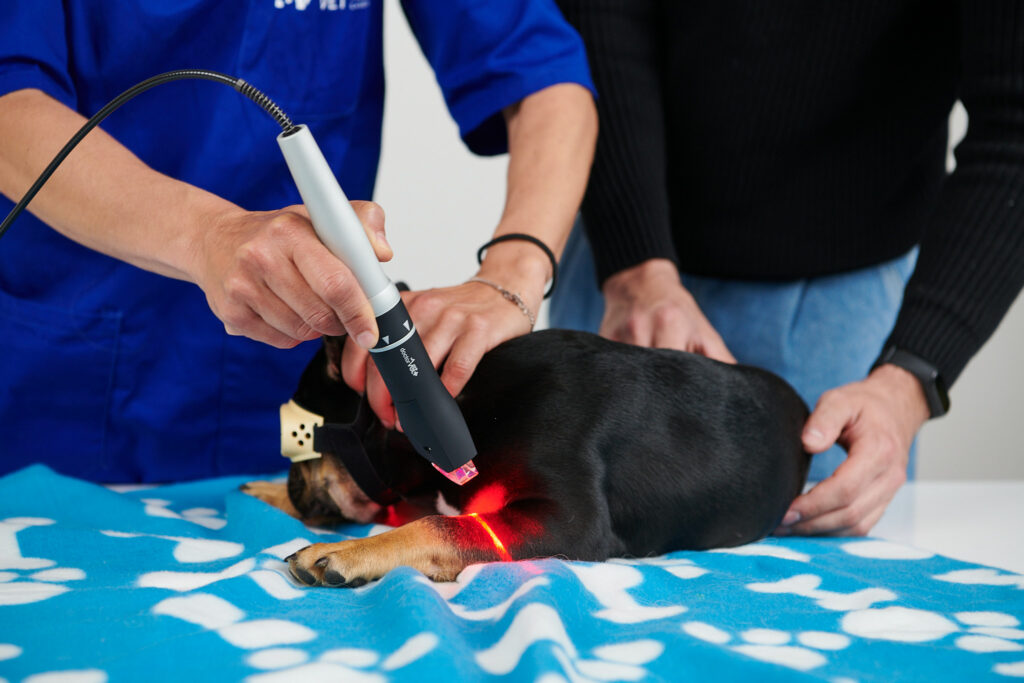
An acceleration in the ossification process has been observed in laser therapy that helps this process in the case of bone fractures or surgeries that require osteotomy. This ossification process takes place due to the effects that occur at a cellular level, with an increase in osteocalcin, collagen, RUNX-2, vascular endothelial growth factors and morphogenetic protein being observed1.
LIGAMENTS AND TENDONS are characterised by consisting of type I collagen with little cellularity. These cells are known as tenocytes and are aligned with the forces of tension. They present very little vascularisation, which is why the repair process is very reduced. The difference between ligaments and tendons is that ligaments present a less parallel alignment, contain less elastin and cells and are more dense and resistant.
In scar tissue, this tissue presents a loss of elastin content. It also loses the more organised structure that presents physiologically.
Laser therapy has shown itself to be useful in the tendon and ligament repair process, with various studies in veterinary medicine having been conducted. This improvement in the repair process is related to the stimulation of collagen synthesis, improving the biomechanical properties of connective tissue2.
HYALINE CARTILAGE is formed of an extra-cellular matrix of proteoglycans and collagen. The cells that form this extra-cellular matrix are chondrocytes. It is an avascular and aneural tissue located at the end of bones.
Because of these characteristics, healing capacity is very limited and depends on the depth of the wound. In wounds with cartilage affectation, a certain repair process occurs through the osmosis of nutrients from the synovial liquid and certain proliferation of fibroblasts.
When there is affectation of the subchondral bone, blood vessels may invade the cartilage. The repaired tissue presents fewer biomechanical characteristics.
It has been observed that laser therapy helps the repair process for this hyaline cartilage by boosting the creation of blood vessels and blood oxygenation, maintaining proteoglycan content, reducing cellular changes and controlling the inflammatory process3.
MUSCLE TISSUE presents different characteristics. Striated muscle is of interest to us from an orthopaedic point of view. One of the characteristics presented by this tissue is its contractile capacity. Formed of myocytes and muscle fibres, cytoplasm consists of perfectly ordered myofibrils. An inflammatory process in this tissue leads to reduced contraction capacity. Furthermore, the repair process for this tissue follows a standard pattern but the creation of large fibrotic tissue can be harmful because it can reduce contraction capacity.
Hence the importance of controlling the inflammatory process. Laser therapy helps to control the entire muscle healing process by increasing or reducing ROS levels and IGF-1 and TGF-β1 growth factors. This homeostasis causes muscular regeneration and reduces the formation of fibrotic tissue4.
In each one of its protocols for musculoskeletal problems, DoctorVethas five stages related to the stimulation of each one of these tissues given that a wound is not only affected by one tissue but rather all the tissues that make up a joint. It also has a stage for the stimulation of microcirculation. This means it can be of assistance in the treatment of various musculoskeletal problems.
BIBLIOGRAPHY
Hosseinpour et al. Molecular impactos of photobiomodulation on bone regeneration: a systematic review. Prog Biophys Mol Biol. 149: 147-159. 2019
Pluim et al. High-power laser therapy improves healing of the equine suspensory branch in a standardized lesion model. Frontiers in vet.sci. Vol. 7 (September). 2020
Felizatti et al. Effects of low-level laser therapy on the organization of articular cartilage in an experimental microcrystalline arthritis model. Lasers in med. Science. 2019
Luo et al. Effects of low-level laser therapy on ROS homeostasis and expression of IGF-1 and TGF-β1 in skeletal muscle during the repair process: Lasers med. Sci (28): 725-734. 2013
Do you want to know more about DoctorVet applications?