Pododermatitis in birds is defined as a degenerative inflammatory process that can cause deep infections, including lesions on tendons and osteomyelitis of the metatarsal bones and phalanges1.
Knowing the anatomical characteristics of this region is essential in understanding the pathology and the seriousness of its effects. The plantar surface is characterised as a rough, uneven surface made up of numerous protuberances (papillae). The pad is formed of connective tissue and fat between the dermis and the tendons. Despite these characteristics, there is very little distance between the surface and the tendons and bones2.
The first signs that this pathology is appearing are ischaemia and necrosis of the soft tissues, leading to the release of inflammatory mediators. This aggravates the lesion and causes thrombosis of small blood vessels.
Inflammation, erythema, scabs and moist lesions can be observed at an initial stage.
Hyperkeratosis appears when the lesion becomes chronic, reducing the epithelium thickness followed by tissue ulceration. This lesion can advance towards the subcutaneous tissue, potentially causing secondary infections. The most common micro-organisms in the Psittacidae family are Staphylococcus spp3.
If the situation is left untreated, it may lead to osteomyelitis, tendinopathies and arthritis. At this stage, the prognosis is unfavourable as it leads to systemic pathologies such as endocarditis and polyarthritis4.
A I to V classification scale for pododermatitis has been established in birds of prey: I = the most minor of pododermatitis; to V = with the most serious prognosis)5.
Knowing the factors that lead to the pododermatitis is essential and may include excess weight (considering that larger species have a greater predisposition)3, perch characteristics, lack of activity, nutrition (such as hypervitaminosis A in Psittacidae and a lack of biotin in turkeys) and lack of hygiene in their cages2.
A clinical history and examination of these patients is essential for diagnosis. Full analyses should be performed to ensure there are no accompanying diseases aggravating the situation or infections present. The seriousness of the lesions may also need to be assessed with imaging techniques2.
In terms of treatment, topical treatments, bandages and systemic treatments may be required based on the seriousness of the lesions, as well as surgery to excise the necrotic tissue. Furthermore, the environment of the patient must be improved in case the causes stem from poor handling.
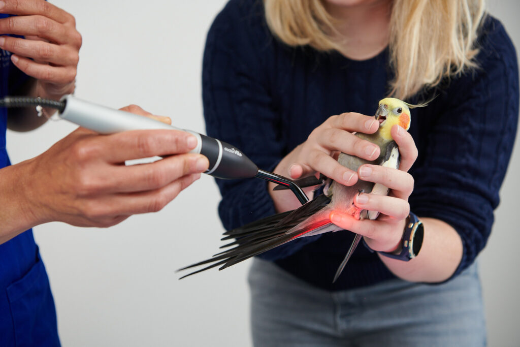
Laser therapy is recommended for pododermatitis in various exotic species2. This therapy can help stimulate microcirculation6, the stimulation of fibroblasts7, the control of inflammation8 and help combat possible infections9.
DoctorVet has specific protocols for birds. To treat pododermatitis, we recommend using the zoom treatment head, the scanning application technique and non-contact mode. The recommended protocols may vary depending on the seriousness of the lesions and may be a combination of inflammation with open wound, subcutaneous (depending on the seriousness of the lesion) and/or infection.
BIBLIOGRAPHY
McCluggage D. Avian medicine and surgery. 1st Ed. Saunders: 830-832, 1997.
Blair J. Bumblefoot: a comparision of clinical presentation and treatment of pododermatitis in rabbits, rodents, and birds. Vet Clin North Am Exot Anim Pract, 16 (3): 715-735, 2013.
Bauck L. Avian medicine and surgery. 1st Ed. Saunders: 554-556, 1997.
Stoute ST. et al. Mycotic pododermatitis and mycotic pneumonia in commercial turkey poults in Northern California. J Vet Diagn Invest, 21(4): 554-557, 2009.
Remple JD. Raptor bumblefoot: a new treatment technique. 154-160, 1993
Karu et al. Cellular effects of low power laser therapy can be mediated by nitric oxide. Lasers in surg. Medicine 36: 307-314, 2005.
Kara N. et al. Laser therapy induces increased viability and proliferation in isolated fibroblast cells. Wounds, 32(3): 69-73, 2020.
Prianti et al. Low-level laser therapy (LLLT) reduces the COX-2 mRNA expression in both subplantar and total brain tissues in the model peripheral inflammation induced by administration of carrageenan. Lasers Med.Sci. 2014.
Silva DC et al. Low level laser therapy (AlGaInP) applied at 5J/cm2 reduces the proliferation of Staphylococcus aureus MRSA in infected wounds and intact skin of rats. An Bras Dermatol, 88(1): 50-55, 2013.
Do you want to know more about DoctorVet applications?