Hip dysplasia: which dog breeds are most affected?
Hip dysplasia is a non-congenital hereditary condition occurring in large and giant breeds. It is also related to environmental factors, such as excess high-energy diet, high activity during development and excess weight. Subluxation or luxation of the femoral head occurs due to abnormal development of the coxofemoral joint. Articular degenerative disease or osteoarthritis (OA) is a secondary complication.
The Orthopaedic Foundation for Animals includes Labradors, Retrievers, Golden Retrievers, German Shepherds, Rottweilers, Bulldogs, Pugs and Saint Bernards in this category1. No greater predisposition occurs between genders. However, patients sterilised at an early age show a greater predisposition to suffer from this disease2. This castration at an early age refers to patients between 5.5 and 12 months.
The symptoms may vary depending on the degree of dysplasia, potentially appearing at an early age or in adults as a result of degenerative changes occurring as a side effect of coxofemoral joint instability.
The symptoms include: intolerance to exercise, avoiding stairs, difficulty lying down, hip pain to the touch, limps of varying severity, walking with both rear limbs at the same time (“rabbit-like”), discomfort when extending the hip, gluteal musculature atrophy, iliopsoas muscle discomfort and crepitation (which indicates OA in the joint).
Diagnosis is based on a correct anamnesis, as well as a correct examination of the patient, which includes the Ortolani test, the Barlow test and the Barden while the animal is anaesthetised in order to assess the degree of laxitude in the coxofemoral joint.
X-rays can also be taken, which require correct positioning. The degree of subluxation or luxation that appears, the flattening of the acetabulum and the presence of degenerative signs (reduced joint space, sclerosis, osteophytes, changes in the shape of the femoral acetabulum, head and neck) may be observed.
Therapeutic and surgical treatments
Treatment may vary:
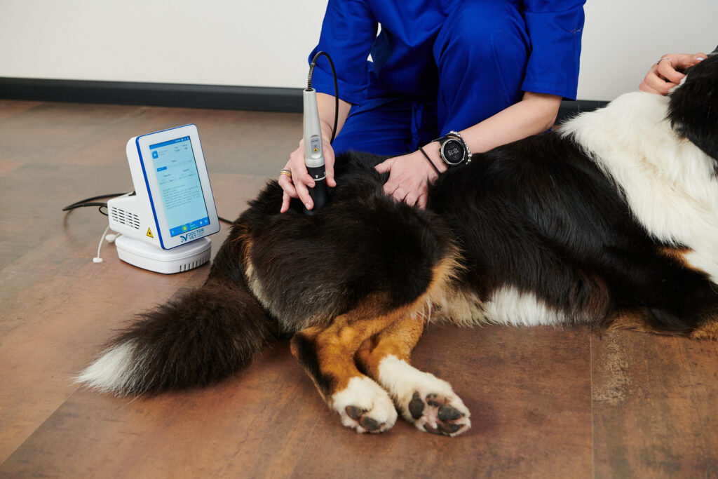
Conservative treatment: multi-modal therapy that may include weight control, correct nutrition, use of AINE and analgesics, rehabilitation (laser therapy is among the physical agents recommended) and the use of chondroprotectors and neutrocytes.
Surgical treatment: various surgical techniques exist, including double and triple pelvic osteotomy, pubic symphysiodesis and hip prosthesis (hip arthroplasty can also be considered in cases of significant pain but this is not satisfactory in animals of a large size). The various surgical techniques are chosen according to the characteristics of the patient3.
Laser therapy can help both conservative and surgical treatments4. In the former, the initial goal is to control the pain and inflammation, and then to help maintain good muscular condition by stimulating microcirculation and creating a biostimulant effect in the articular cartilage. If a correct therapeutic protocol can be established, it will be possible to reduce pharmacological treatment and therefore any potential side effects.
In surgical treatment, the goal is to reduce the pain and inflammation after surgery. The goal then shifts to reducing recovery time through correct ossification.
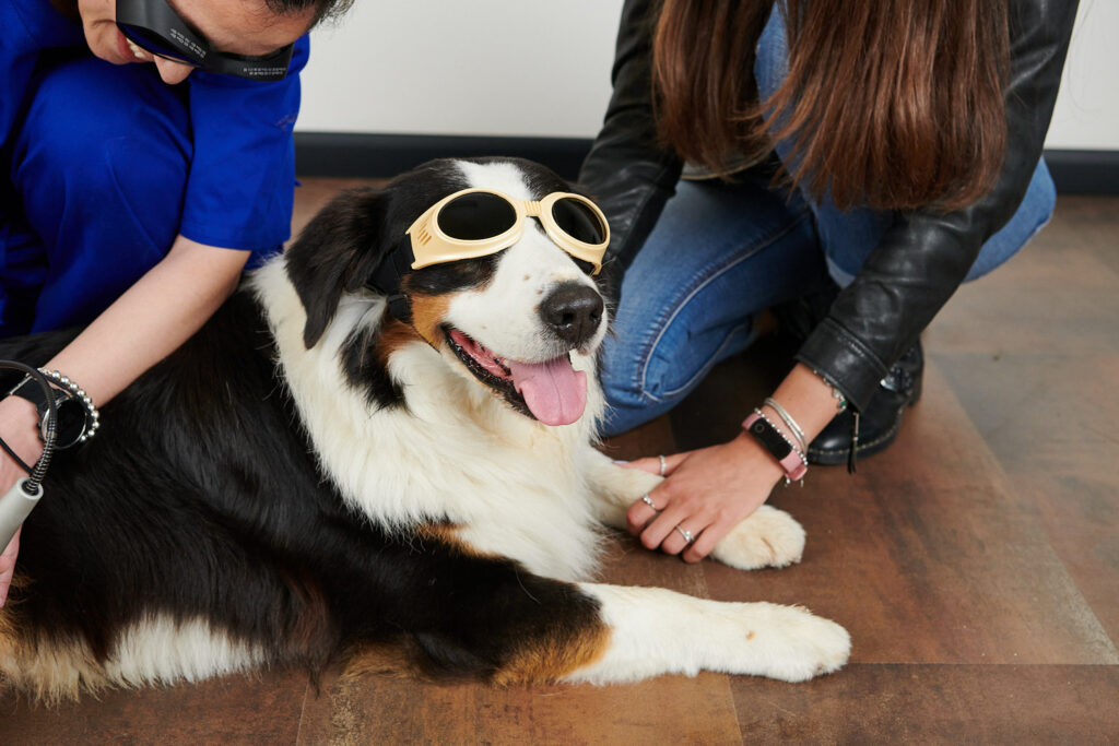
DoctorVetoffers a specific protocol for hips capable of treating hip dysplasia. The ideal solution for this pathology is to treat using a hand tool and avoid excessive pressure, especially if the patient presents limb atrophy, as this can be uncomfortable. If contact cannot be tolerated, the scanner hand tool can be used in non-contact mode.
The specific hip protocol can be combined with the inflammation and general pain protocol depending on the symptoms shown by the patient. Treatment can begin with three sessions per week until the effects of photobiomodulation are seen. Maintenance sessions can be then performed, which may vary from one treatment every 15 days to one treatment every 4-5 weeks.
BIBLIOGRAPHY
Orthopedic Foundation for Animals. Hip Dysplasia Statistics by disease.
Spain et al. Long-term risk and benefits of early-age gonadectomy in dogs. J Am Vet Med Assoc, 224: 380-7. 2004
Piermattei et al. Brinker, Piermattei, and Flo´s handbook of small animal orthopaedics and fracture repair. Pág: 416-511. 4th edit. 2006
Prydie et al. Practical physiotherapy for small animal practice. Chap: 11. Ed. Wiley. 2015
Do you want to know more about DoctorVet applications?